X-Tomography Synchrotron
X-ray computed tomography (CT) is a non-destructive technique for visualizing features within solid objects and for obtaining digital information about their geometry and 3-dimensional properties.
Synchrotron X-Ray Tomography provides a high-quality X-ray energy source to acquire the three-dimensional internal structure of real objects non-destructively and with high spatial resolution from micrometers to nanometers. This allows detailed microstructural analysis of many different materials such as small engineering components.
Synchrotron X-ray tomography is based on detecting the attenuation or phase shift of the beam passing through a sample. While X-ray scans measure images for one direction of the sample, CT scans measure images for many different angular positions. This results in a set of projections, which can be used to reconstruct two-dimensional layers or slices through the object. By stacking these slices together, the structure can be visualized in three dimensions.

Synchrotron-generated X-ray beams provide the following advantages:
Micro-CT is a 3D imaging technique that uses X-rays to look inside an object, slice by slice. Micro-CT, also known as micro-tomography or computed tomography, is similar to hospital CT or CAT scan imaging but on a small scale with greatly increased resolution. Samples can be imaged with pixel sizes as small as 100 nanometers, and objects can be scanned as large as 200 mm in diameter.
The Micro-CT scanner captures a series of 2D planar X-ray images and reconstructs the data into 2D horizontal slices. These slices can be further processed into 3D models and even printed as 3D physical objects for analysis. With a 2D X-ray system, you can see through an object, but with the power of a 3D micro-CT system, you can see inside an object and reveal its internal features. It provides volumetric information about the microstructure, not the structure.
X-rays are generated in the X-ray source, transmitted through the sample, and recorded by the X-ray detector as a 2D projected image. The sample is then rotated a fraction of a degree on the rotation stage and another X-ray image is taken. This step is repeated through rotation 180 degrees (or sometimes 360 degrees, depending on the sample type). The series of X-ray projection images are then computed into cross-sectional images through a computational process called Reconstruction. These slices can be analyzed, further processed into 3D models, rendered into movies, printed into 3D physical objects, and more.

Non-destructive testing (NDT) means that the sample or pattern being scanned is not altered or destroyed during the test or during test preparation. This allows the sample to be preserved for historical record, retested at a later date, used in another test, or put into final production. Some other techniques require staining, cutting, or coating the sample, which may affect the sample's structure, continued usefulness, or use in future studies. There are several techniques that allow samples to be imaged in their natural state, including optical microscopy, laser scanning, spectroscopic imaging, and other visible observations, etc. Micro-CT is one such technique in which most of the samples under study are scanned in an unchanged state.
Micro-CT provides high-resolution 3D imaging information that cannot be obtained with any other non-destructive technology. It can be used to study the internal structure of both material and biological samples without having to cut the sample, preserve the sample or sample for future studies. Quantitative information obtained from micro-CT scans can only be obtained from 3D images, and 3D digital models created from micro-CT virtual slices allow scientists to measure any parameter for comparison. compared in before and after studies.
These unique features of micro-CT scanning allow scientists to examine the sample's morphology and study characteristics such as: porosity, bone structure/thickness, volume fraction, defect analysis, density, grain size, voids, fiber orientation, etc. Use micro-CT to study bones, teeth, tissues/organs, composites, medical devices, batteries, and more.
CT scan images have many applications: in medicine, in Materials Science as well as in Life Sciences.
Micro-CT scans are 3D X-ray images, using the same method as medical CT (or CAT) scans, but micro-CT is on a much smaller scale with greatly increased resolution. Medical CT scanning was introduced as a tool for medical imaging in the 1970s; CT scans (or computed tomography) are limited to 1 millimeter resolution, which provides enough detail for clinical use. For materials science and small animal imaging, much higher resolution was needed, and micro-CT scanning was introduced in the 1980s. Micro-CT scanners can operate at one micron, one thousandth millimeter and smaller.
Simply put, in vivo (Latin for in life) is the scanning of living specimens and ex vivo (Latin for living) usually refers to things that once existed or samples taken from something alive. As for micro-CT, in vivo generally refers to systems that scan rats and mice and in some cases rabbits, while ex vivo systems typically handle the rest of the applications.

With in vivo micro-CT instruments, because the animals are still alive, longitudinal studies can be performed to measure the effects of drugs, diet, hormones, and other treatments on tumor; bone growth and quality; body mass; and other applications in the same topic. This can reduce the number of animals needed for a study.
Ex vivo microsurgical devices typically handle residual applications, including endpoint studies of specific regions of the animal being resected (lungs, bones, tumors, implants, grafts, etc.) , biomaterials research, implantation in large animals, materials research, compression research, and more. Ex-vivo micro-CT devices allow for higher spatial resolution, longer scan times (due to dose to the sample of concern), better signal to noise ratio and thus better imaging . Ex vivo systems are commonly used for most applications outside of living animals.
Nanotomography (nano-CT) is similar to micro-CT and medical CT scanning but at a resolution in nanometers instead of microns or millimeters. Sigray's Nano-CT uses a nano-focusing X-ray source to capture 2D images during 180 or 360 degree rotation of the sample. Advanced software is then used to turn those images into 2D cross-sections or slices through the sample. These cross-sections give the researcher the opportunity to look inside the sample without having to cut it open. The smaller the focal point of the X-ray source, the higher the resolution that can be achieved on the sample scan. Nano-CT is important for observing details as best as possible without destroying the sample.
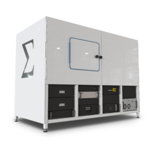
Main Feature:
Applications:


